
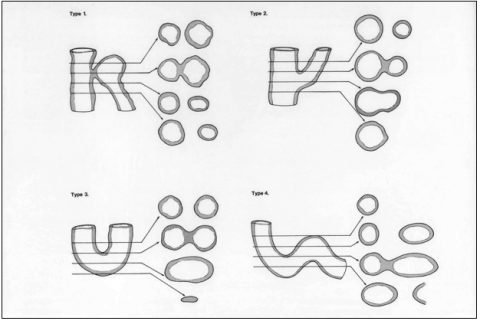
 The villous tree of the human placenta displays a complex 3-dimensional topology. Not only do the villi branch repeatedly, but the longer villi also follow sinuous courses. As a result, a variety of villous profiles can be seen in any single histological section, and it is often impossible to appreciate how one profile relates to another; are they two separate villi or the same villus curving around? In particular, tangential sections through the trophoblast layer can generate the appearance of trophoblastic bridges linking adjacent villi, or syncytial knots protruding from the surface (see figure) [1, 2]. The only way to be sure of what one is looking at is to follow the structure through serial sections.
Images derived from serial sections from two placentas are available here for anyone who would like to follow structures in 3-D and appreciate this point. The two sets are derived from normal term placentas that were delivered by cesarean section. Immediately after delivery the placentas were immersed intact in 10% formol saline for 4 weeks. Full-thickness blocks of tissue, from the chorionic to the basal plates, were then removed, and embedded in paraffin wax. Two stacks of 20 serial sections were cut from two placentas. Sections were cut at 7 µm, stained with haematoxylin and eosin, and scanned using a x20 objective on a Hamamatsu NanoZoomer scanner. The scanner captures the entire contents of a microscope slide into a single image, and then compiles the image so that it can be viewed at different magnifications and moved around like a real microscope slide. This is called virtual digital microscopy.
The whole slide images can be accessed by clicking each image file name. This then opens up the image in the NDP server window. You do not need any special software to use the image browser. Navigation and zooming is intuitive and can be done either using your mouse or the control panel at the top left hand side of the browser window.
Index You can go to the slide index page by clicking the Back button from your web browser.
Zooming You can move around the slide by clicking and holding at the same time as moving your mouse/trackpad. In the bottom right hand corner of the image there is always a small inset window showing you whereabouts you are in the slide when you are zooming at high power. Zooming is achieved by clicking on the magnification tool in the control panel and select the desired virtual objective lens (see figure). You can always return to the slide overview by using the overview button on the control panel.
Measurements The NDP software allows you to make linear or area measurements using the annotation tool. Click on the "pencil" menu and select "Linear Measure" or "Rectangle Area". You can now draw a line or an area on the image and obtain a measurement in micrometers or square micrometers. You can export/save image by clicking the floppy disk button on the control panel.
Slide images of Placental serial section A & B
[1] Burton GJ. Intervillous bridges in the mature human placenta; syncytial fusion or section artifacts? J Anat 1986;145:12-23.
[2] Cantle SJ, Kaufmann P, Luckhardt M, Schweikhart G. Interpretation of syncytial sprouts and bridges in the human placenta. Placenta 1987;8:221-34.