Boyd Collection is an archival collection of human embryonic material held at the University of Cambridge. The specimens were collected in the 1950s and 1960s by Professor JD Boyd, Professor of Anatomy at the University. Professor Boyd was a leading embryologist who wrote the classic monographs 'Human Embryology' (Hamilton, Boyd and Mossman) and 'The Human Placenta' (Boyd and Hamilton). Because of his research interests local pathologists sent Professor Boyd specimens for review. The collection is comprised solely of histological sections, and no tissue blocks remain.
The placental samples fall into two categories; placenta-in-situ specimens and isolated placental specimens. Few clinical details are available from which to assess the normality of the samples. The placenta-in-situ specimens often demonstrate low-lying placentas, and may have resulted from hysterectomy for ante-partum haemorrhage. The isolated placental samples are likely to have been obtained from spontaneous miscarriages.
Researchers who wish to view the slides should contact the Centre's Administrator.
We are aiming to scan representative sections of the placenta-in-situ specimens and place them on the web as a resource for researchers.
We have started with the larger sections, and will add others in due course. The sections have been scanned on a high resolution flat-bed scanner, and both low and high resolution files are available. The high resolution files have been tiled using Zoomify, allowing viewers to move around the section.
The approximate gestational ages dated from the last menstrual period are based on records of the crown-rump length. The size of each tissue section is given for reference purposes. The quality of fixation is variable, and in some cases the chorion laeve has separated from the decidua parietalis due to shrinkage.
When sections can be identified as having been used in 'The Human Placenta' or when they are clearly from the same block, reference is made to the appropriate figure number.
The images can be downloaded and used for illustrative and teaching purposes, but if they are used in publications acknowledgement should be made to the Loke Centre for Trophoblast Research.
|
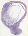
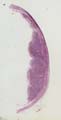
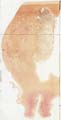
CR length 20 mm. Approximate gestational age 8.5 weeks. Section size 7.0 cm x 5.8 cm. Stain; H & E. View – Download |
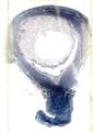
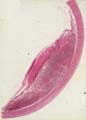
CR length 55 mm. Approximate gestational age 12 weeks. Section size 9.5 cm x 7.0 cm. Stain; Chrome. View – Download |
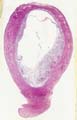
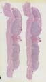
CR length 60 mm. Approximate gestational age 12.5 weeks. Section size 11.0 cm x 7.5 cm. Stain; Mallory's trichrome. View – Download |
|
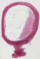
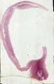
CR length 73 mm. Approximate gestational age 14 weeks. Section size 11.0 cm x 6.5 cm. Stain; H & E. View – Download |
CR length 90 mm. Approximate gestational age 15.5 weeks. Section size 12.5 cm x 7.5 cm. Stain; H & E. Fig. 112 in 'The Human Placenta'. View – Download |
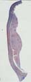
CR length 140 mm. Estimated about 20.5 weeks. Section size 11.0 cm x 4.0 cm. Stain; H & E. Fig. 120 in 'The Human Placenta'. Labelled as a good example of 'trophoblast giant cells in the decidua'. View – Download |
|
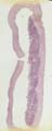
CR length 145 mm. Approximate gestational age 21 weeks. Section size 8.5 cm x 2.5 cm. Stain; H & E. View – Download |
CR length 205 mm. Approximate gestational age 27 weeks. Section size 11.0 cm x 3.0 cm. Stain; H & E. Fig. 132 in 'The Human Placenta'. View – Download |
CR length 210 mm. Approximate gestational age 27.5 weeks. Section size 14.0 cm x 8.5 cm. Stain; H & E. Fig. 134 in 'The Human Placenta'. View – Download |
|
CR length 220 mm. Approximate gestational age 28.5 weeks. Section size 15 cm x 7.5 cm. Stain; Van Gieson. View – Download |
CR length 240 mm. Approximate gestational age 30.5 weeks. Section size 16.0 cm x 4.0 cm. Stain; trichrome. View – Download |
CR length 260 mm. Approximate gestational age 32.5 weeks. Section size 14.5 cm x 4.5 cm. Stain; H & E. Fig. 137 in 'The Human Placenta'. View – Download |